Tired all the time

73 year old presents with general tiredness

Differential Diagnosis

What are the differential diagnosis for general tiredness?

Show answer

A differential diagnosis for tiredness is quite broad, therefore using a framework may help in adding structure and uncovering your knowledge.

We'll use the surgical sieve 'VINDICATE':

  • Vascular - Systemic vasculitis or heart failure
  • Inflammatory / Infection - Chronic infection such as an abscess or endocarditis
  • Neoplastic - Must be ruled out especially in older patients
  • Denegerative
  • Iatrogenic - Medications or poorly controlled diseases
  • Autoimmune - Inflammatory Bowel Disease (IBD) leading to malabsorption
  • Traumatic - Sleep apnoea (not really trauma! But certainly a mechanical cause of chronic tiredness)
  • Endocrine - Diabetes, Hypothyroidism

Remember the functional and psychiatric conditions that may present as tiredness such as depression and chronic fatigue syndrome.

Taking a history

If we want to rule out a gastrointestinal cause, what are the key questions?

Show answer

Obviously because this case sits within a gastrointestinal section we'll focus on those parts of the history, but patients don't present as easily as this!

When taking a GI history it can be thought of as:

  • Pain - Any abdominal pain? Site and onset are of most interest but remember to take a full SOCRATES pain history (you can read about this in the case Chest Pain)
  • "Ins" - What is the patient's appetite? Are they able to keep fluids down? Is the pain related to eating?
  • "Outs" - Vomiting? If so, what colour? Black and red indicate upper GI bleeds. Opening bowels? Absolute constipation points towards obstruction, whilst diarrhoea leans more towards inflammatory or infective. Any change in bowel habit could be a sign of malignancy.
  • Jaundice - Often easy to forget about asking this but can be a good question to rule in / rule out pathologies.
  • B-symptoms - Fever, weight loss and night sweats - these should pretty much roll off your tongue as a standard set of 'red flag' symptoms for any given history.

History

Robert Fence is a 73 year old retired builder who has been growing progressively more tired for the last 6 months. He has reported no other symptoms of note apart from an occasional discomfort in his abdomen. He has not been bloated nor has he had any bloody stool, and has never had a positive Faecal Occult Blood Test. He has noticed his stools have become looser in the last 4 months.

He has lost weight of 3.3kg without changing his diet or exercise regime, he is not overly thirsty and doesn't report any change to his skin colour.

Mr Fence has no past medical history of note and has self reportedly been in good health for as long as he can remember. He has no allergies and is on no regular medication. he is an ex-smoker of 10 pack years and drinks moderate amounts of alcohol. He lives at home with his wife and is fully independant. He is concerned about the progressive nature of his tiredness.

History

What are the causes of altered bowel habit?

Show answer

Again when thinking of a differential diagnosis try and use a framework to help pin your ideas to.

When dealing with tube-like structures one technique is to think of causes anatomically; in the lumen, in the bowel wall, outside the bowel wall.

  • In the lumen - Diarrhoea is commonly due to infection such as viruses (rotavirus, norovirus) and bacteria (E-coli - food poisoning, C.diff - if recent antibiotic use). Or prescribed medications.
  • In the wall - Inflammatory Bowel Disease (bloody diarrhoea), diverticulitis, malignancy (diarrhoea or constipation), Coeliac disease.
  • Outside the wall - Hyperthyroidism.

Histology

What are the normal layers of the bowel wall?

Show answer

Different pathological processes affect different layers, so make sure you're familiar with them as you may be asked by an inquisitive consultant on a ward round.

  1. Lumen
  2. Mucosa - epithelium, basement membrane, lamina propria
  3. Submucosa
  4. Muscularis propria

Key Concept: Layers of the Gastrointestinal tract

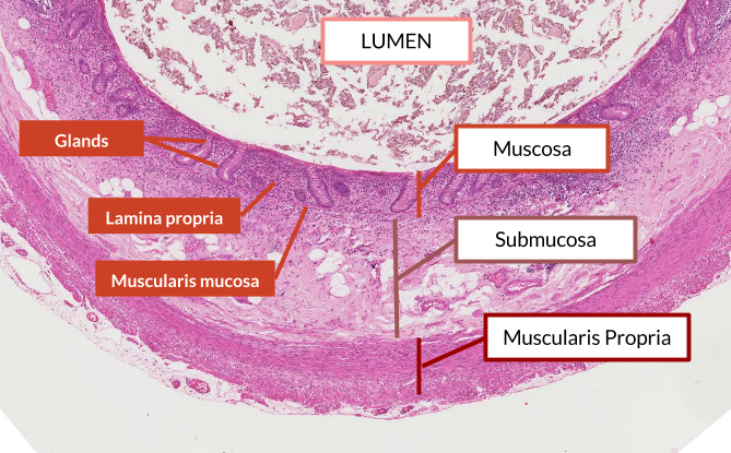
The normal layers of the GI tract can be difficult to remember, so we've created a short video to talk you through.

Below is how the layers appear under a microscope.

Examination

General inspection: Patient comfortable at rest.

Obs:

Abdominal examination: Mr Fence had a pale conjunctiva. Abdomen was soft, non-tender with no mass and no organomegaly. Digital rectal examination was normal.

Abdominal examination

Mr Fence's examination findings point towards anaemia. What are the different signs of anaemia?

Show answer

A very basic routine for any examination would be general inspection, hands, face and finally the system you are examining. Using this approach you may find the following in someone with anaemia:

  • Koilonychia - "spoon" shaped nails in iron-deficiency anaemia (think spoons that are made of iron!).
  • Tachycardia & tachypnoea - a conpensatory mechanism from lack of oxygen carriage seen in all types of anaemia.
  • Pale conjunctiva - due to lack of haemaglobin colour in iron-deficiency anaemia, as seen in our patient in this case.
  • Angular stomatitis - cracks in the corner of the lips due to iron-deficiency anaemia or low B12.
  • Glossitis - a large, sore tongue due to B12 deficiency.

Investigations

Mr Fence has a set of bloods taken.

FBCValueNormal range
WBC12.844.5 - 13.5 x 10^9 /L
RBC4.094.0 - 6.0 x 10^12 /L
Hb90 *130-150 g/L
MCV78 *90 - 120 fL

* denotes abnormal result

U&E: NAD

LFT: NAD

Glucose: 4.9 mmol/L (Normal range: 4.4 - 6.1 mmol/L).

Data interpretation

What do these blood results indicate? What would we see in folate & B12 deficiencies?

Show answer

The results show a microcytic anaemia (small cells, but normal in number).

  • Commonly related to iron deficiency and blood loss.
  • Consider also anaemia of chronic disease, thallasaemia and sideroblastic anaemia (bonus points if you thought of any of these!).

Blood loss in the GI tract is either:

  • Visible - for bleeding to be seen it must be a lower GI bleed (or a massive upper GI bleed!); haemorrhoids, inflammatory bowel disease, left-sided bowel malignancy, prolapse and ulceration, diverticulosis.
  • Occult - blood would be mixed in with the stool if it is from higher up in the GI tract; gastric / duodenal ulcer, inflammatory bowel disease (Crohn's), right sided malignancy.

Key Concept: Anaemia

The best way to picture anaemia is to think of what goes into making a red blood cell.

Haemoglobin is a protein with iron molecules at it's core. Red blood cells are packed full of haemoglobin, so it makes sense that if iron stores are low then each red blood cell becomes smaller and this manifests as a microcytic anaemia.

Folate and B12 are building blocks of DNA and so are needed to replicate, therefore deficiencies in these will decrease the number of cells, meaning each cell ends up having more haemoglobin and is therefore macrocytic (larger cells).

Guidelines

What should be the next step for this patient? Is there a guideline that could assist?

Show answer

Urgent 2 week referral for colonoscopy for any of the following:

  • Age 40+ with new bowel symptoms
  • Rectal bleeding and persistent change in bowel habit for > 6 weeks
  • Iron-deficiency anaemia
  • Palpable mass (abdominal or rectal).

So which of these does our patient have?

Worryingly he has 3 out of the 5; remember any one of these would merit an endoscopy.

You may find it useful to read the full NICE guidline.

Demographics

What would be your top differentials if the patient was 25 years old?

Show answer

We'd worry more about an inflammatory or infective process, although remember that certain genetic conditions predispose to early onset bowel cancer. Bonus points for naming some.

Key Concept: Inflammatory Bowel Disease

Inflammatory Bowel Disease (IBD) is a chronic inflammatory autoimmune condition that has two major sub-types: Ulcerative Colitis and Crohn's disease.

The main two symptoms are bleeding per rectum and diarrhoea.

Other symptoms/signs that are included in IBD include:

  • Low grade fever
  • Malaise (a term for 'generally unwell')
  • Abdominal pain
  • Loss of weight without intention

Note: all these symptoms would have been uncovered with our abdominal history we covered above.

Ulcerative Colitis and Crohn's disease are closely related and both have similar treatments (immunosuppression medication), so why do you need to know the differences? Well because it always comes up in exams.

Ulcerative ColitisCrohn's
GeneticsDRB-1DQ4 DR7
AetiologyTwo peaks: 20-25; 50-60 M=FOne peak: 40-60 F>M
PathologyStarts at rectum, pseudopolyps due to recovering ulcers, mucosa only affected, loss of goblet cells, thin wallsAnywhere from mouth to anus, full thickness granulomatous inflammation, cobblestone appearance with skipped lesions, most common site is terminal ileum
SequelaeSignficant increase in colorectal cancer riskFistulas, strictures and bowel obstruction, abscess, mile increase in colorectal cancer

Management

Mr Fence went to hospital for a colonoscopy which revealed several polyps in the ascending and transverse colon and a large mass in the caecum of the colon. This was biopsied and found to be malignant.

Pathology

What is a polyp? Does it need to be removed?

Show answer

Bowel polyps are small growths on the inner lining of the colon (large bowel) or rectum. They are common, affecting 15-20% of the UK population, and don't usually cause symptoms.

Bowel polyps are not usually cancerous, although if they're discovered they'll need to be removed, as some will eventually turn into cancer if left untreated.

You can read more about polyps at NHS choices.

Key Concept: Dysplasia

It is important to understand the link between dysplasia and malignancy.

Histology of a polyp on a stalk (pedunculated) areas 1, 2 &3 from bottom up representing normal dysplastic and neoplastic regions.

Concluding the case

After Mr Fence had his colonoscopy he was informed that it was a cancerous growth. The fact that Mr Fence's cancer was confined to his colon (that it had not metastasised), him being a fit and healthy man for his age, it was advised that he have surgery to remove the caecum, the ascending colon and the hepatic flexure.

To learn more about types of operations done please visit: Cancer Research UK.

Self-assessment

What is the order of the layers of the gastrointestinal wall?

Different pathological processes affect different layers, so make sure you're familiar with the normal structure.

What does microcytic hypochromic anaemia mean?

Think about what goes into making a red blood cell.

Which of these complications are most likely to arise from Ulcerative Colitis?

You can reason this question out by knowing two things, that Crohn's disease can be inflammation anywhere from mouth to anus and that it is full thickness inflammation. Small bowel obstruction for example is much more likely from Crohn's strictures as the lumen of the small intestine is much thinner (around half the size) of the large bowel.

A single mutation in which gene is responsible for the dominantly heritable familial adenomatous polyposis colorectal cancer?

All of these markers are involved somehow in cancer, do you know what cancer they're involved in?

True or False: Polyps are very likely to be cancerous?

A simple way of remembering is that 10% polyps will turn cancerous within 10 years.

What is the most common Histology for a colorectal cancer cell?

Remember it is glandular epithelium in the GI tract because of it's secretory nature, therefore an 'adenocarcinoma'.

What is the name of the lymphoid tissue in the large bowel?

This is important as it can be a site of lymphoma.

Which of the following symptoms would require an urgent 2 week referral for colonoscopy?

The correct NICE guidelines are covered above.

What is the definition of Diverticulitis?

Diverticulosis is merely the presence of outpouching, which can lead to diverticulitis - inflammation in these pouches.

------- is the name of the process used to tell how far along a cancer is.

Be sure that you know concepts like this.

Answered: 2/10 Score: 50%

Objectives

Wow! You made it all the way to the end. These are the objectives you will have picked up along the way.

  1. Describe the Aetioogy of colorectal carcinoma
  2. Describe the morphology of colorectal cancer
  3. Describe the normal histology of the GI tract
  4. Describe the pathological consequences of colorectal carcinoma
  5. Describe the pathological change of the histology in neoplasia of the colon
  6. Define Inflammatory Bowel Disease
  7. Indicate the difference in pathology between Crohn's disease and Ulcerative Colitis
  8. Describe additional manifestations of inflammatory bowel disease